Introduction
|
|
Recently, it has been reported that medical students and trainee doctors perceive neurology as one of the more difficult disciplines of medicine [1] [2] [3] [4] [5] [6] [7] [8]
[9]. Furthermore, it has been found that medical students and physician residents feel less confident in the field of neurology than other specialties of medicine [2]. The fear of the neural sciences and clinical neurology, termed "neurophobia", is a common problem among students and trainee doctors worldwide [1] [2] [3] [4] [5] [6] [7] [8][9]. Traditionally, a difficult topic to grasp in clinical neurology is brainstem vascular syndromes [2] [10] [11]
[12]. This article provides a brief overview of the etiology of brainstem vascular syndromes and a simplified approach for medical students and trainee doctors to localize the vascular territory affected. We discuss the rule of 4 and then provide a basic overview of each of the major vascular syndromes emphasizing clinical manifestations, distinguishing features, and relevant neuroanatomy for each syndrome. The aim of this article is to be used as a supplement to neuroscience textbooks used by medical students and healthcare professionals.
|
The Rule of 4
|
|
The rule of 4 is a simplified method to localize brainstem vascular syndromes specific to a vascular territory. Utilizing the rule of 4 as well as distinguishing clinical signs specific to each vascular syndrome, the vascular territory affected can be quickly and accurately localized. There are 4 basic rules to this schema [13]:
First Rule: There are 4 midline structures that begin with the letter M
Second Rule: There are 4 lateral structures that begin with the letter S
Third Rule: There are 4 cranial nerves below the pons, 4 in the pons, and 4 above the pons
Fourth Rule: There are 4 midline cranial nerve motor nuclei
Within each of the 4 rules are 4 additional guidelines necessary to understand in order to localize the site of vascular occlusion [13].
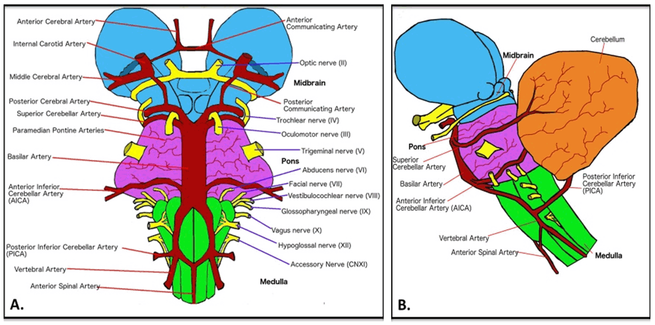
Third rule: There are 4 cranial nerves (CN) below the pons, 4 in the pons, 4 above the pons (Figure 1):
- Medulla: glossopharyngeal, vagus, spinal accessory, hypoglossal (CN 9–12, respectively)
- Pons: trigeminal, abducens, facial, vestibulocochlear (CN 5–8, respectively)
- Midbrain: oculomotor, trochlear (CN 3–4, respectively)
Fourth rule: There are 4 midline motor nuclei: The nuclei of cranial nerves 3, 4, 6, and 12:
|
| Cursor on image to zoom/Click text to open image |
| |
Figure 1: Brainstem vasculature and emerging cranial nerves (A) Ventral view of the brainstem, (B) Lateral view of the brainstem. The brainstem is organized into three divisions. Caudal to cranial they are the medulla oblongata, pons, and midbrain. At the level of the medulla, four cranial nerves emerge including the glossopharyngeal, vagus, accessory, and hypoglossal nerves. At the level of the pons, four cranial nerves emerge including the trigeminal, abducens, facial, and vestibulocochlear nerves. At the level of the midbrain, two cranial nerves emerge including the oculomotor and trochlear nerves. The trochlear nerve is the only nerve to exit via the dorsal surface of the brainstem as well as immediately decussate. The olfactory and optic nerves emerge above the level of the midbrain.
|
|
Vascular Syndromes of The Medulla Oblongata
|
|
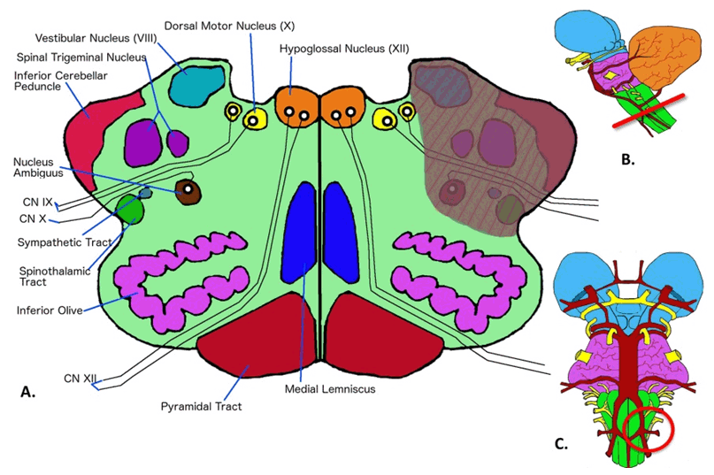
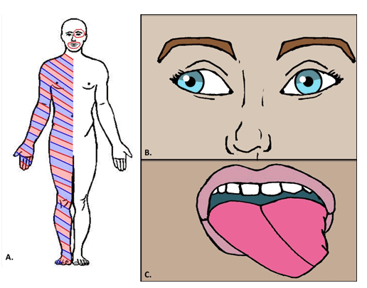
Lateral Medullary Syndrome
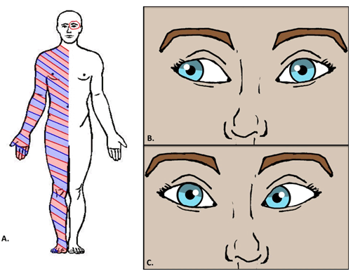
Lateral medullary syndrome, also known as Wallenberg syndrome, is usually due to infarction of the posterior inferior cerebellar artery (PICA) (Figure 2) [14] [15]. The posterior inferior cerebellar artery supplies the lateral territory of the medulla. With the lateral medulla affected, the second rule of 4 applies to this vascular syndrome. Patients with lateral medullary syndrome present with (Figure 3):
- loss of pain and temperature of the contralateral arm and leg (spinothalamic tract)
- ataxia of the ipsilateral arm and leg (spinocerebellar tract)
- horner's syndrome of the ipsilateral eye (sympathetic pathway)
- loss of pain and temperature of the ipsilateral face (sensory nucleus of trigeminal nerve)
Distinguishing features of lateral medullary syndrome
The third rule indicates that cranial nerves 9–12 may be affected in medullary infarcts. The fourth rule excludes cranial nerve 12 from being affected in this particular syndrome, as cranial nerve 12 is a midline structure. This leaves cranial nerves 9, 10, and 11 as possible cranial nerves affected by a lateral medullary infarct. Cranial nerves 9 and 10 are affected in this particular syndrome as cranial nerve 11 is usually not affected in medullary infarcts. Differentiating features specific to lateral medullary syndrome are loss of function of cranial nerves 9 and 10 (glossopharyngeal and vagus nerve, respectively) resulting in hoarseness and dysphagia [14] [15]. A simple pneumonic to remember these distinguishing features as well as the artery affected is "Never pick a (PICA) horse (hoarseness) that cannot eat (dysphagia) [16].
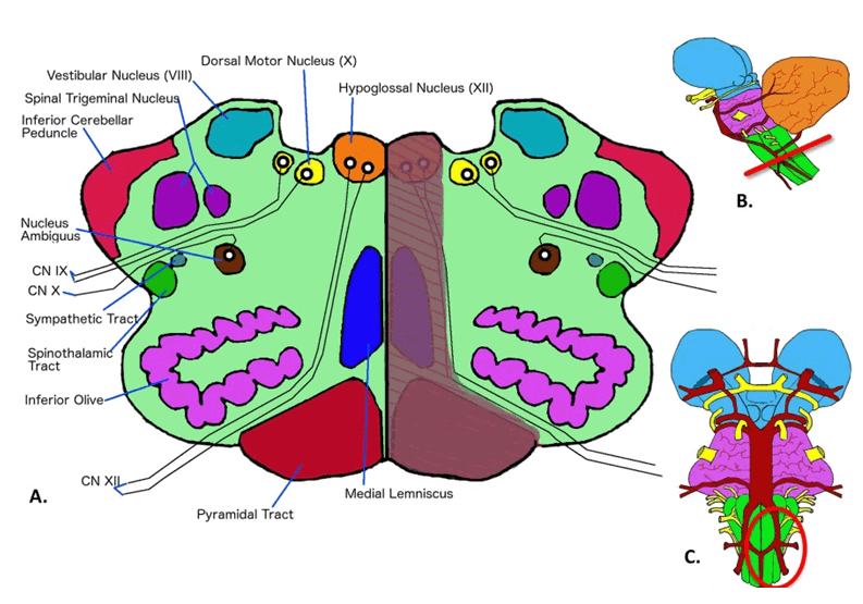
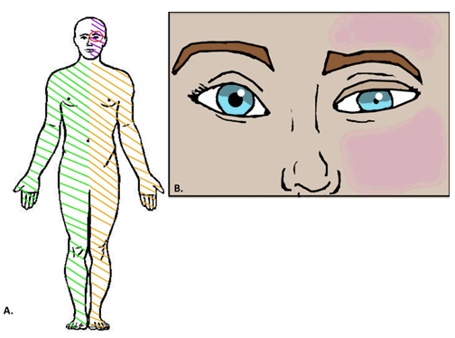
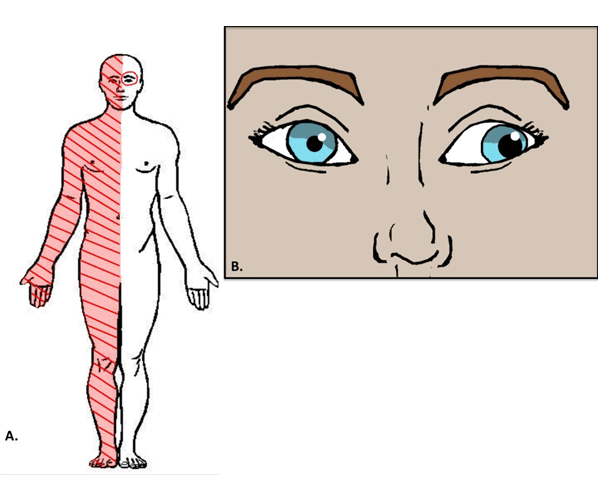
Medial Medullary Syndrome
Medial medullary syndrome, also known as Dejerine syndrome, is most commonly due to infarction of the vertebral artery (Figure 4) [15] [17]. The vertebral artery supplies the medial territory of the medulla. As the medial medulla is affected, the first rule of 4 applies to this vascular syndrome. Patients with medial medullary syndrome present with (Figure 5):
- weakness of the contralateral arm and leg (corticospinal tract)
- loss of vibration and proprioception of the contralateral arm and leg (medial lemniscus)
- internuclear ophthalmoplegia of the ipsilateral eye (medial longitudinal fasciculus)
- loss of function of midline ipsilateral cranial nerve 12
Distinguishing features of medial medullary syndrome
The third rule indicates that cranial nerves 9–12 may be affected in medullary infarcts. The fourth rule indicates that cranial nerve 12 is the only cranial nerve affected in this syndrome because it is the only cranial nerve that arises midline and from the medulla. A differentiating feature specific to medial medullary syndrome is loss of function of cranial nerve 12 (hypoglossal nerve) resulting in deviation of the hypoglossal nerve to the ipsilateral side (side of the infarction) [15] [17]. A simple pneumonic to remember that the tongue deviated to the ipsilateral side when the hypoglossal nerve is affected is "lick your wounds" – referring to the tongue deviating towards the side of the "wound" (infarct) [16].
|
|
|
|
|
|
|
|
|
|
Vascular Syndromes of The Pons
|
|
Lateral Pontine Syndrome
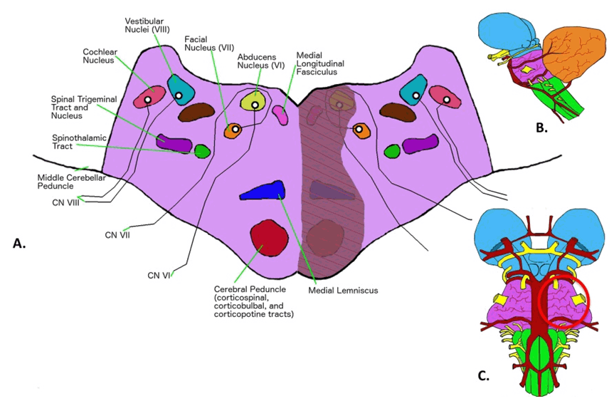
Lateral pontine syndrome usually results from infarction of the anterior inferior cerebellar artery (AICA) (Figure 6) [15]. The anterior inferior cerebellar artery supplies the lateral territory of the pons. With the lateral pons affected, the second rule of 4 applies to this vascular syndrome. Patients with lateral pontine syndrome present very similar to patients with lateral medullary syndrome, except for different distinguishing cranial nerve features. Patients with lateral pontine syndrome present with (Figure 7):
- loss of pain and temperature of the contralateral arm and leg (spinothalamic tract)
- ataxia of the ipsilateral arm and leg (spinocerebellar tract)
- horner's syndrome of the ipsilateral eye (sympathetic pathway)
- loss of pain and temperature of the ipsilateral face (sensory nucleus of trigeminal nerve)
Distinguishing features of lateral pontine syndrome
The third rule indicates that cranial nerves 5–8 may be affected in pontine infarcts. The fourth rule excludes cranial nerve 6 from being affected in this particular syndrome, as cranial nerve 6 is a midline structure. This leaves cranial nerves 5, 7, and 8 as possible cranial nerves affected by a lateral pontine infarct. A differentiating feature specific to lateral pontine syndrome is loss of function of cranial nerve 7 (facial nerve) resulting in facial paralysis [15]. A simple pneumonic to remember that facial nucleus effects are specific to AICA lesions is "facial droop means AICA is pooped" [16].
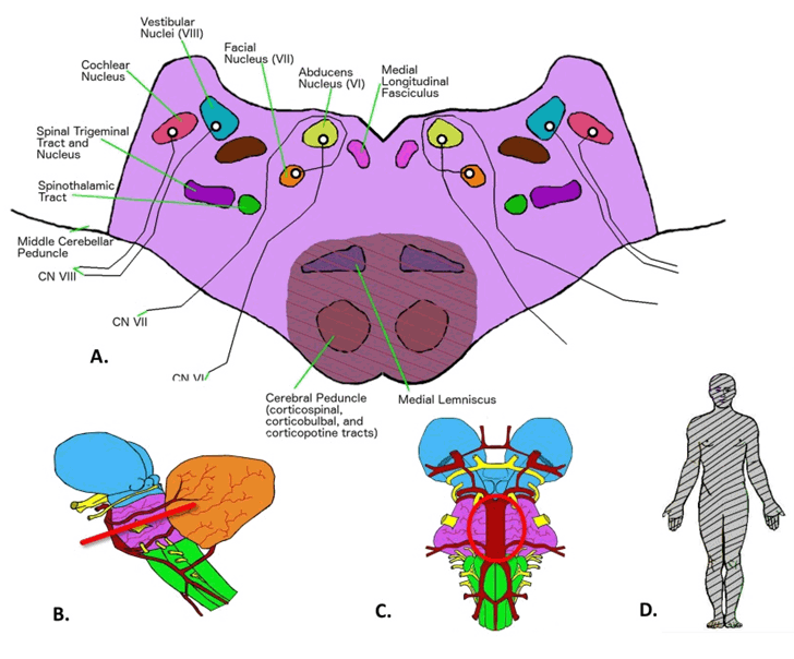
Medial Pontine Syndrome
Medial pontine syndrome most commonly results from infarction of the paramedian branches of the basilar artery (Figure 8) [15] [18]. The paramedian branches of the basilar artery supply the medial territory of the pons. As the medial pons is affected, the first rule of 4 applies to this vascular syndrome. Patients with medial pontine syndrome present very similar to the patients with medial medullary syndrome, except for different distinguishing cranial nerve features. Patients with medial pontine syndrome present with (Figure 9):
- weakness of the contralateral arm and leg (corticospinal tract)
- loss of vibration and proprioception of the contralateral arm and leg (medial lemniscus)
- internuclear ophthalmoplegia of the ipsilateral eye (medial longitudinal fasciculus)
- loss of function of midline ipsilateral cranial nerve 6
Distinguishing features of medial pontine syndrome
The third rule indicates that cranial nerves 5–8 may be affected in pontine infarcts. The fourth rule indicates that cranial nerve 6 is the major cranial nerve affected in this syndrome, as cranial nerve 6 is a midline structure. A differentiating feature specific to medial pontine syndrome is loss of function of cranial nerve 6 (abducens nerve) resulting in strabismus [15] [18]. Strabismus is characterized as ipsilateral paralysis of the lateral rectus muscle (the eye that is affected will look inferior and towards the nose).
Ventral Pontine Syndrome
Ventral pontine syndrome, also known as cerebromedullospinal disconnection, or locked-in syndrome, is caused by infarction of the basilar artery (Figure 10). The basilar artery supplies the pons. The first and second rule of 4 applies to this vascular syndrome. Ventral pontine syndrome is easy to identify as a collection of bilateral long tract signs (motor and sensory) sometimes supplemented by signs of cranial nerves 5–8 dysfunction. This results in the quadriplegia and aphasia. However, patients with this syndrome usually do not exhibit paralysis of the eyes. Individuals with locked-in syndrome may be able to communicate by moving their eyes or blinking [15] [19].
|
|
|
|
|
|
|
|
|
|
|
|
|
Vascular Syndromes of The Midbrain
|
|
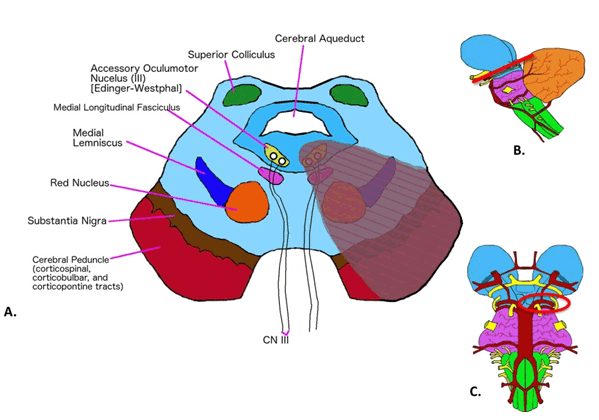
Medial Midbrain Syndrome
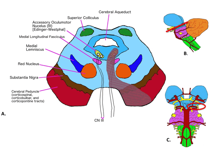
Medial midbrain syndrome, also known as Weber's syndrome, is usually due to infarction of the penetrating branches of the posterior cerebral artery (Figure 11) [15] [20]. The penetrating branches of the posterior cerebral artery supply the midbrain [15] [20]. Unlike the medulla and pons, vascular syndromes of the midbrain do not exactly follow the rule of 4. Occlusion of these branches in this syndrome results in (Figure 12):
- ipsilateral eye "down and out" with dilation, and an unresponsive pupil (oculomotor nerve)
- weakness of the contralateral face (corticobulbar tract)
- weakness of the contralateral arm and leg (corticospinal tract)
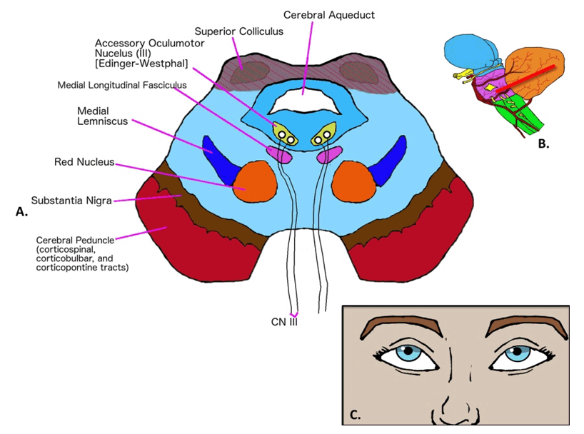
Lateral Midbrain Syndrome
Lateral midbrain syndrome, also known as Benedikt syndrome, is most commonly due to penetrating branches of the posterior cerebral artery (Figure 13) [15] [20]. The penetrating branches of the posterior cerebral artery supply the midbrain [15] [20]. Occlusion of these branches in this syndrome results in (Figure 14):
- ipsilateral eye "down and out" with dilation, and an unresponsive pupil (oculomotor nerve)
- ataxia of the contralateral body (red nucleus)
- tremor (dentatorubrothalamic tract)
Dorsal Midbrain Syndrome
Dorsal midbrain syndrome, also known as Parinaud's syndrome and vertical gaze palsy, is most commonly caused by a pinealoma, which compresses the superior colliculus at the rostral interstitial nucleus of medial longitudinal fasciculus (Figure 15) [15]. This syndrome can also result from multiple sclerosis and stroke of the upper brainstem. It should be noted that case reports in the medical literature have demonstrated that occlusion of the posterior thalamo-subthalamic paramedian artery can also cause this syndrome [21][22]. Nevertheless, the classic etiology of Parinaud's syndrome, caused by a pinealoma, results in:
- paralysis of upward gaze (rostral interstitial nucleus of medical longitudinal fasciculus)
- collier's sign (retraction of the eyelids)
|
|
|
|
|
|
|
|
|
|
|
|
|
Conclusion
|
|
In summary, if one can remember the rule of 4 and the distinguishing cranial nerve feature(s) of each vascular syndrome then one will be able to identify the presenting syndrome and localize the stroke to a vascular territory.
|
References
|
-
Lim EC, Seet RC. Demystifying neurology: Preventing 'neurophobia' among medical students. Nat Clin Pract Neurol 2008 Aug;4(8):462–3.
[CrossRef]
[Pubmed]

-
Zinchuk AV, Flanagan EP, Tubridy NJ, Miller WA, McCullough LD. Attitudes of US medical trainees towards neurology education: "Neurophobia" - a global issue. BMC Med Educ 2010 Jun 23;10:49.
[CrossRef]
[Pubmed]
-
McColgan P, McKeown PP, Selai C, Doherty-Allan R, McCarron MO. Educational interventions in neurology: A comprehensive systematic review–reply to letter. Eur J Neurol 2013 Oct;20(10):e123.
[CrossRef]
[Pubmed]
-
Pakpoor J, Handel AE, Disanto G, Davenport RJ, Giovannoni G, Ramagopalan SV; Association of British Neurologists. National survey of UK medical students on the perception of neurology. BMC Med Educ 2014 Oct 21;14:225.
[CrossRef]
[Pubmed]
-
Flanagan E, Walsh C, Tubridy N. 'Neurophobia'–attitudes of medical students and doctors in Ireland to neurological teaching. Eur J Neurol 2007 Oct;14(10):1109–12.
[CrossRef]
[Pubmed]
-
Matthias AT, Nagasingha P, Ranasinghe P, Gunatilake SB. Neurophobia among medical students and non-specialist doctors in Sri Lanka. BMC Med Educ 2013 Dec 9;13:164.
[CrossRef]
[Pubmed]
-
Youssef FF. Neurophobia and its implications: Evidence from a Caribbean medical school. BMC Med Educ 2009 Jul 1;9:39.
[CrossRef]
[Pubmed]
-
McGee J, Maghzi AH, Minagar A. Neurophobia: A global and under-recognized phenomenon. Clin Neurol Neurosurg 2014 Jul;122:iii–iv.
[CrossRef]
[Pubmed]
-
Ramos RL, Cuoco JA, Guercio E, Levitan T. Quantitative Description of Medical Student Interest in Neurology and Psychiatry. J Am Osteopath Assoc 2016 Jul 1;116(7):462–71.
[CrossRef]
[Pubmed]
-
Nicholl DJ, Appleton JP. Clinical neurology: why this still matters in the 21st century. J Neurol Neurosurg Psychiatry 2015 Feb;86(2):229–33.
[CrossRef]
[Pubmed]
-
Nham B. Graded exposure to neurophobia: Stopping it affect another generation of students. Australian Medical Student Journal 2012;3(1).
-
Eid E, Dastan S, Heckmann JG. Acute dizziness in rural practice: Proposal of a diagnostic procedure. J Neurosci Rural Pract 2015 Apr-Jun;6(2):272–6.
[CrossRef]
[Pubmed]
-
Gates P. The rule of 4 of the brainstem: A simplified method for understanding brainstem anatomy and brainstem vascular syndromes for the non-neurologist. Intern Med J 2005 Apr;35(4):263–6.
[CrossRef]
[Pubmed]
-
Shetty SR, Anusha R, Thomas PS, Babu SG. Wallenberg's syndrome. J Neurosci Rural Pract 2012 Jan;3(1):100–2.
[CrossRef]
[Pubmed]
-
Longo DL, Fauci AS, Kasper DL, Hauser SL, Jameson JL, Loscalzo J. eds. Harrison's Principles of Internal Medicine. 18ed. New York, NY: McGraw Hill; 2012. p. 3288–93.
-
Le T, Bhushan V, Sochart M. First Aid for the USMLE Step 1. New York, NY: McGraw Hill; 2016. p. 46.
-
Kim K, Lee HS, Jung YH, et al. Mechanism of medullary infarction based on arterial territory involvement. J Clin Neurol 2012 Jun;8(2):116–22.
[CrossRef]
[Pubmed]
-
Ruhland JL, van Kan PL. Medial pontine hemorrhagic stroke. Phys Ther 2003 Jun;83(6):552–66.
[Pubmed]
-
Laureys S, Pellas F, Van Eeckhout P, et al. The locked-in syndrome: what is it like to be conscious but paralyzed and voiceless? Prog Brain Res 2005;150:495–511.
[CrossRef]
[Pubmed]
-
Bailey BJ, Johnson JT, Newlands SD. Head & Neck Surgery–Otolaryngology. 4ed. Philadelphia, PA: Lippincott Williams & Wilkins; 2006. p. 119.
-
Serino J, Martins J, Páris L, Duarte A, Ribeiro I. Parinaud's syndrome due to an unilateral vascular ischemic lesion. Int Ophthalmol 2015 Apr;35(2):275–9.
[CrossRef]
[Pubmed]
-
Clark JM, Albers GW. Vertical gaze palsies from medial thalamic infarctions without midbrain involvement. Stroke 1995 Aug;26(8):1467–70.
[Pubmed]
|