|
Case Report
Neurodegenerative lesion of tumefactive multiple sclerosis demonstrates the fluorescence radiation from 5-aminolevulinic acid-induced protoporphyrin IX
1 MD, Director, Department of Neurosurgery, Hokuto Hospital, Obihiro, Hokkaido, Japan
Address correspondence to:
Akira Tempaku
7-5, Inada-cho-kisen, Obihiro, Hokkaido 080-0833,
Japan
Message to Corresponding Author
Article ID: 100025N06AT2025
Access full text article on other devices

Access PDF of article on other devices


How to cite this article
Tempaku A. Neurodegenerative lesion of tumefactive multiple sclerosis demonstrates the fluorescence radiation from 5-aminolevulinic acid-induced protoporphyrin IX. Edorium J Neurol 2025;10(2):16–20.ABSTRACT
Tumefactive multiple sclerosis (tMS) reveals imaging findings in the central nervous system alongside extensive surrounding edematous changes. The nature and etiology of the disease are often unknown. In addition, differentiation from a primary intracranial tumor is often difficult based on imaging studies alone. Therefore, surgical resection and pathological examination are often required to obtain a definitive diagnosis. A 48-year-old woman attended the clinic complaining of headaches. A ring-shaped, contrast-enhanced lesion with perifocal edema in magnetic resonance imaging was noted in the right occipital lobe. She underwent craniotomy, and pathological examination revealed a diagnosis of tMS. Intracranial findings at the time of surgical operation showed excitatory light positivity for 5-aminolevulinic acid-induced protoporphyrin IX (PPIX). In tMS lesions, the author observed PPIX accumulation, indicative of mitochondrial dysfunction. It can be hypothesized that mitochondrial metabolic disturbance would result in inhibition of heme synthesis. The hypothesis that the decrease in adenosine triphosphate (ATP) production due to heme depletion is the underlying cause of the energy deficit is one that merits further investigation. This finding of mitochondrial dysfunction accompanied by the decrement of PPIX metabolization might be important for the etiologic analysis of tMS.
Keywords: 5-Aminolevulinic acid Fluorescence, Protoporphyrin IX, Tumefactive multiple sclerosis
INTRODUCTION
Tumefactive demyelinating lesions (TDL) are defined as solitary intracranial demyelinating lesions more than 2 cm in diameter [1],[2],[3],[4],[5]. Neuroradiology shows low signal on T1-weighted magnetic resonance image (MRI), high signal on T2-weighted MRI, and ring enhancement effect on T1 of gadolinium (Gd) contrast administration [6]. Compared to the size of the lesion, it is characterized by a less compressive effect on the surrounding brain [7]. The incidence of TDL is rare at 0.3/100,000/year, more common in women, and the mean age of onset has been reported to be 37 years [3]. Tumefactive multiple lesions have been reported to be the most common form of TDL in Japan. Among them, tumefactive multiple sclerosis (tMS) is a rare subtype of multiple sclerosis (MS) with TDL, occurring at a frequency of 0.1–0.2% of MS [8]. Focal demyelinating degeneration is the predominant cause. Depending on the site of onset, it can lead to motor paralysis, sensory deficits, higher brain dysfunction, epilepsy, and other disorders. However, the pathogenesis and pathophysiology of tMS remain unclear [9].
In some cases, the diagnosis may be difficult to distinguish from a brain tumor. A definitive diagnosis is usually made by the pathological examination following to the surgical removal of the lesion [7],[10],[11]. Here reports on intraoperative findings that may help to elucidate the pathophysiology of the disease.
CASE REPORT
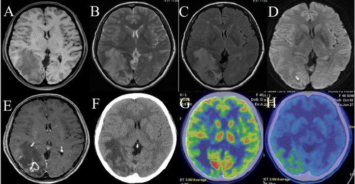
A 48-year-old woman presented to the hospital with a headache for several days. She was not accompanied by impaired consciousness (scoring 15 points on the Glasgow Coma Scale: 4 points for eye movement, 5 points for verbal response, and 6 points for motor response), hemiplegia (scoring 5 points for all rims on the Manual Muscle Testing scale), visual field disorder, or higher brain dysfunction (scoring 28 points on the Japanese version of the Mini-Mental State Examination, and 43 seconds for part A and 84 seconds for part B of the Japanese version of the Trail Making Test). Seizure or seizure-like clinical episode was not observed. An MRI scan of the head revealed a 3 cm diameter mass lesion in the right occipital lobe with T1 iso, T2 hypo, flow attenuated inversion recovery (FLAIR) hypo and diffusion-weighted image (DWI) hyper (Figure 1). Gadolinium-enhanced magnetic resonance imaging showed ring-like enhancement (Figure 1). Computed tomography (CT) scan showed iso density, which was surrounded with low density area (Figure 1). Perfusion CT showed that cerebral blood volume (CBV) was unchanged, but cerebral blood flow (CBF) was slightly decreased (Figure 1). Positron emission tomography (PET) scan showed negative in 5-fluorodeoxyglucose (FDG) accumulation (Figure 1) and weak positive in methionine accumulation (Figure 1).
The patient had a mild fever of 37.0 °C and an elevated white blood cell count of 13,470/µL, but there was no evidence of liver or kidney dysfunction. There were no electrolyte or coagulation abnormalities, and the C-reactive protein (CRP) test was negative. Tumor markers were also negative. No significant abnormal findings were noted in the examination of the cerebral spinal fluid (CSF). Subsequently, the patient also complained of disturbance to her left visual field (including transient visual hallucinations and photosensitivity).
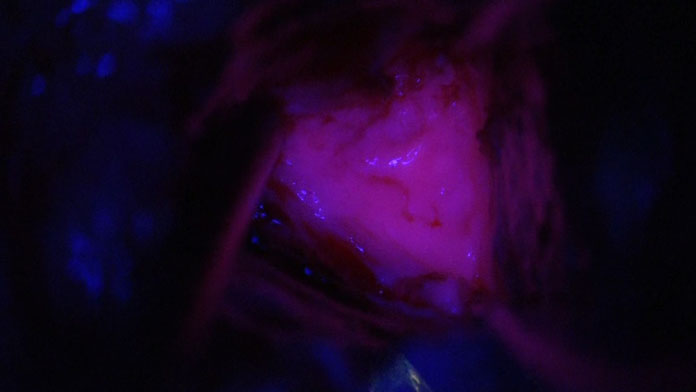
Based on imaging studies, glioma, brain abscess, and metastatic tumor were listed as differential diagnoses. However, these diagnoses could not be confirmed through physical and radiological imaging examinations alone. A pathological diagnosis was deemed necessary, so a craniotomy was performed under general anesthesia one month after the initial diagnosis. Using optical navigation system (StealthStation7, Medtronic, Dublin, Ireland) in the park bench position, the lesion was located and the parenchymal portion of the mass was removed. Intraoperative observation under excitation light illumination was found to be positive for 5-aminolevulinic acid (Ala)-induced protoporphyrin IX (PPIX) fluorescence. In order to ascertain the excitation light, the core of the lesion was irradiated with ultraviolet light at a wavelength of 400 nm (Blue 400) under the microscopic view of KINEVO 900 (Carl Zeiss Meditec, Jena, Germany). As illustrated in Figure 2, excitation light induced by 5Ala was observed to be pink in color.
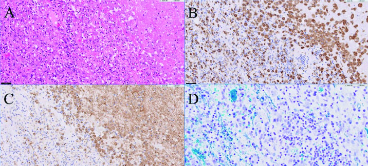
Pathological examination revealed a necrotic lesion with low cell density and inflammatory cell infiltration. Inflammatory cells were predominantly cluster of differentiation (CD) 68-positive histiocytes (Figure 3). Myelin basic protein staining and Kluver–Barrera staining show the demyelinated status (Figure 3). Grocott’s stain showed no evidence of fungal or protozoan infection. Histologically, tumefactive demyelinating lesions (TDL) were suspected. Additional serological tests showed negative in anti-myeline oligodendrocyte glycoprotein (MOG) and anti-aquaporin (AQP) 4 antibodies. A diagnosis of tumefactive multiple sclerosis was made three months after the initial diagnosis.
Treatment with steroids was initiated once the diagnosis had been confirmed. Methylprednisolone sodium succinate was given intravenously at 1 g per day for the first three days. Prednisolone was then taken orally for the following two weeks at 30 mg per day, decreasing gradually. The patient’s residual lesions, including edematous changes, were resolved for 20 days. The patient was discharged home without any neurological complications at five months after the initial diagnosis.
DISCUSSION
Tumefactive MS is predominantly a focal, central demyelinating lesion, with imaging findings similar to high-grade gliomas [2],[3],[4],[5]. It is characterized by extensive surrounding edema on T2 and FLAIR sequences, and ring-like enhancement on gadolinium-enhanced MRI. However, it usually lack some imaging findings that are typically associated with high-grade gliomas, such as weak methionine accumulation and increased vascularity and blood flow to the lesion on angiography. There may also be a lack of arteriovenous (AV) shunt. There is extensive edema relative to the size of the lesion. Some patients have extensive edema but lack the characteristics of malignancy. These include mild cerebral swelling and peri-brain compression [4]. A diagnosis is often difficult to confirm using preoperative imaging studies alone. The final diagnosis is made by removing and examining the tumor under pathological study.
As far as the author was able to ascertain, no reports focusing on the presence or absence of 5Ala induced PPIX fluorescence were found. However, there are some case reports describing intraoperative fluorescence as either weakly positive [12],[13] or negative [14]. No definitive findings have yet been established.
Five Ala is the enzyme substrate of 5-aminolevulinate dehydratase, which is widely present in various organisms. Five Ala is taken up and degraded in the mitochondria to produce adenosine triphosphate (ATP), which is used as an energy substrate [15],[16],[17],[18],[19],[20],[21],[22]. Protoporphyrin IX, an intermediate product of this degradation process, fluoresces when exposed to excitation light [20],[21],[22],[23]. Since the function of mitochondria is reduced in tumor cells, the intermediate metabolite of 5Ala, PPIX, accumulates. This technique has been applied to the surgical removal of tumors, including glioma, and has contributed to improve therapeutic outcomes [24],[25],[26].
An increase in the fluorescence of 5Ala induced PPIX was observed in tMS tissue. This was thought to be caused by an increase in 5Ala uptake associated with demyelination as well as the accumulation of PPIX due to decreased 5Ala metabolism associated with mitochondrial damage [27].
In rats, it has been reported that loss of mitochondrial function causes demyelination [21]. Under normal conditions, the migration of mitochondria into the axon of the demyelinated area contributes to protection from neurodegeneration [23]. However, in demyelinating diseases such as MS, dysfunction of mitochondria within neurons is observed, which leads to neurodegeneration [28].
The positive fluorescence of 5Ala induced PPIX during surgery proves that demyelinating tissues in tMS are accompanied by mitochondrial dysfunction.
CONCLUSION
This report captures the reduction in mitochondrial activity in tMS tissue in vivo using the accumulation of 5Ala induced PPIX as an indicator. Decreased mitochondrial metabolic activity may be the underlying cause of tMS formation. Further studies are needed to analyze the pathogenesis.
REFERENCES
1.
van der Velden M, Bots GT, Endtz LJ. Cranial CT in multiple sclerosis showing a mass effect. Surg Neurol 1979;12(4):307–10.
[Pubmed]

2.
Dagher AP, Smirniotopoulos J. Tumefactive demyelinating lesions. Neuroradiology 1996;38(6):560–5. [CrossRef]
[Pubmed]
3.
Jamroz-Wiśniewska A, Janczarek M, Belniak E, Stelmasiak Z, Bartosik-Psujek H. Tumour-like lesions in multiple sclerosis. Neurol Neurochir Pol 2008;42(2):161–7.
[Pubmed]
4.
Nakayama M, Naganawa S, Ouyang M, Jones KA, Kim J, Capizzano AA, et al. A review of clinical and imaging findings in tumefactive demyelination. AJR Am J Roentgenol 2021;217(1):186–97. [CrossRef]
[Pubmed]
5.
Al Malik YM. Tumefactive demyelinating lesions: A literature review of recent findings. Neurosciences (Riyadh) 2024;29(3):153–60. [CrossRef]
[Pubmed]
6.
Suh CH, Kim HS, Jung SC, Choi CG, Kim SJ. MRI Findings in tumefactive demyelinating lesions: A systematic review and meta-analysis. AJNR Am J Neuroradiol 2018;39(9):1643–9. [CrossRef]
[Pubmed]
7.
Butteriss DJA, Ismail A, Ellison DW, Birchall D. Use of serial proton magnetic resonance spectroscopy to differentiate low grade glioma from tumefactive plaque in a patient with multiple sclerosis. Br J Radiol 2003;76(909):662–5. [CrossRef]
[Pubmed]
8.
Schwartz KM, Erickson BJ, Lucchinetti C. Pattern of T2 hypointensity associated with ring-enhancing brain lesions can help to differentiate pathology. Neuroradiology 2006;48(3):143–9. [CrossRef]
[Pubmed]
9.
Hu W, Lucchinetti CF. The pathological spectrum of CNS inflammatory demyelinating diseases. Semin Immunopathol 2009;31(4):439–53. [CrossRef]
[Pubmed]
10.
Cha S, Pierce S, Knopp EA, Johnson G, Yang C, Ton A, Litt AW, Zagzag D. Dynamic contrast-enhanced T2*-weighted MR imaging of tumefactive demyelinating lesions. AJNR Am J Neuroradiol 2001;22(6):1109–16.
[Pubmed]
11.
Fallah A, Banglawala S, Ebrahim S, Paulseth JE, Jha NK. Case series: Tumefactive demyelinating lesions: A diagnostic challenge. Can J Surg 2010;53(1):69–70.
[Pubmed]
12.
Suzuki S, Oikawa M, Ito T, Sako K, Ozaki Y, Yoshinaga T, et al. Tumefactive multiple sclerosis: A case report. J Hokkaido Brain Res Found 2008;12:61–64.
13.
Ishikawa K, Satoh K, Ito T, Ozaki Y, Asanome T, Yamaguchi Y, et al. A case of tumefactive multiple sclerosis resembling malignant glioma. Jpn J Neurosurg (Tokyo) 2017;26:688–93.
14.
Kuninaka T, Hokama Y, Tokeshi Y, Namihira Y, Ishiuchi S. A case of demyelinating disease with the solitary and gadolinium ring like enhanced lesion in head MRI. Ryukyu Med J 2021;40(1–4):37–46.
15.
Afonso SG, Chinarro S, Muñoz JJ, de Salamanca RE, Batlle AM. Photodynamic and non-photodynamic action of several porphyrins on the activity of some heme-enzymes. J Enzyme Inhib 1990;3(4):303–10. [CrossRef]
[Pubmed]
16.
Navone NM, Polo CF, Frisardi AL, Andrade NE, Battle AM. Heme biosynthesis in human breast cancer— Mimetic “in vitro” studies and some heme enzymic activity levels. Int J Biochem 1990;22(12):1407–11. [CrossRef]
[Pubmed]
17.
Kondo M, Hirota N, Takaoka T, Kajiwara M. Heme-biosynthetic enzyme activities and porphyrin accumulation in normal liver and hepatoma cell lines of rat. Cell Biol Toxicol 1993;9(1):95–105. [CrossRef]
[Pubmed]
18.
Herman MA, Webber J, Fromm D, Kessel D. Hemodynamic effects of 5-aminolevulinic acid in humans. J Photochem Photobiol B 1998;43(1):61–5. [CrossRef]
[Pubmed]
19.
Casas A, Batlle AM, Butler AR, Robertson D, Brown EH, MacRobert A, et al. Comparative effect of ALA derivatives on protoporphyrin IX production in human and rat skin organ cultures. Br J Cancer 1999;80(10):1525–32. [CrossRef]
[Pubmed]
20.
Kajimoto Y, Kuroiwa T. Unsolved problems in 5-aminolevulinic acid based photodynamic diagnosis: Quantification of fluorescence and molecular mechanism of porphyrin accumulation. JJSLSM 2011;32(2):143–8.
21.
Kuramoto T, Kuwamura M, Tokuda S, Izawa T, Nakane Y, Kitada K, et al. A mutation in the gene encoding mitochondrial Mg²+ channel MRS2 results in demyelination in the rat. PLoS Genet 2011;7(1):e1001262. [CrossRef]
[Pubmed]
22.
Takahashi K. The various applications of 5-aminolevurinic acid (ALA). J Arid Land Stud 2018;28(2):66-72.
23.
Licht-Mayer S, Campbell GR, Canizares M, Mehta AR, Gane AB, McGill K, et al. Enhanced axonal response of mitochondria to demyelination offers neuroprotection: Implications for multiple sclerosis. Acta Neuropathol 2020;140(2):143–67. [CrossRef]
[Pubmed]
24.
Stummer W, Pichlmeier U, Meinel T, Wiestler OD, Zanella F, Reulen HJ; ALA-Glioma Study Group. Fluorescence-guided surgery with 5-aminolevulinic acid for resection of malignant glioma: A randomised controlled multicentre phase III trial. Lancet Oncol 2006;7(5):392–401. [CrossRef]
[Pubmed]
25.
Pichlmeier U, Bink A, Schackert G, Stummer W; ALA Glioma Study Group. Resection and survival in glioblastoma multiforme: An RTOG recursive partitioning analysis of ALA study patients. Neuro Oncol 2008;10(6):1025–34. [CrossRef]
[Pubmed]
26.
Yagi R, Kawabata S, Fukumura M, Omura N, Hiramatsu R, Kameda M, et al. Is Intraoperative 5-ALA PDD effective for brain tumors other than malignant gliomas!? 5-Ala PDD and preoperative image analysis in metastatic brain tumors. JJSLSM 2023;44(2):178–85. [CrossRef]
27.
Novotny A, Stummer W. 5-Aminolevurinic acid and blood-brain barrier – A review. Medical Laser Application 2003;18(1):36–40. [CrossRef]
28.
Campbell GR, Worrall JT, Mahad DJ. The central role of mitochondria in axonal degeneration in multiple sclerosis. Mult Scler 2014;20(14):1806–13. [CrossRef]
[Pubmed]
SUPPORTING INFORMATION
Acknowledgments
The author thanks Dr. Eriko Aimono and Dr. Hiroshi Nishihara for their pathological studies and the medical staff of Hokuto Hospital for supporting clinical treatment and care for the patient.
Author ContributionsAkira Tempaku - Conception of the work, Design of the work, Acquisition of data, Analysis of data, Drafting the work, Revising the work critically for important intellectual content, Final approval of the version to be published, Agree to be accountable for all aspects of the work in ensuring that questions related to the accuracy or integrity of any part of the work are appropriately investigated and resolved.
Guaranter of SubmissionThe corresponding author is the guarantor of submission.
Source of SupportNone
Consent StatementWritten informed consent was obtained from the patient for publication of this article.
Data AvailabilityAll relevant data are within the paper and its Supporting Information files.
Conflict of InterestAuthor declares no conflict of interest.
Copyright© 2025 Akira Tempaku. This article is distributed under the terms of Creative Commons Attribution License which permits unrestricted use, distribution and reproduction in any medium provided the original author(s) and original publisher are properly credited. Please see the copyright policy on the journal website for more information.